
Translate this page into:
Spontaneous subcutaneous emphysema in a male toddler in a health facility in Nasarawa state: A case report
*Corresponding author: Surajudeen Oyeleke Bello, Department of Paediatrics, Dalhatu Araf Specialist Hospital, Lafia, Nasarawa, Nigeria. surajudeenbello4@gmail.com
-
Received: ,
Accepted: ,
How to cite this article: Bello SO, Umejiaku S, Ogunkunle TO, Afolabi OF, Yakubu AA. Spontaneous subcutaneous emphysema in a male toddler in a health facility in Nasarawa state: A case report. J Pan Afr Thorac Soc 2021;2(1):53-5.
Abstract
Background:
Spontaneous subcutaneous emphysema (SSE) is a clinical condition in which air escapes into the subcutaneous tissues. It is a rare complication of childhood pneumonia and often occurs with pneumothorax and/or pneumomediastinum. Although the sight of a child with SSE could be frightening, it is mostly benign requiring in most cases supportive care. We report a case of SSE complicating pneumonia in an 18-month-old toddler that was managed conservatively.
Case Report:
An 18-months-old toddler was admitted with a 5-day history of progressive swelling of the face, scalp, upper limbs, and trunk. He was referred from a peripheral hospital where he was admitted for 5 days with pneumonia and had received antibiotics, intravenous fluid, and oxygen therapy. Clinical evaluation revealed extensive subcutaneous emphysema and right-sided pneumothorax. Antibiotics were optimized and the patient was provided supportive care and monitored for 7 days with complete resolution of the emphysema.
Conclusion:
SSE could complicate childhood pneumonia but it has a benign course. Effective treatment of underlying pneumonia alongside supportive care will achieve complete resolution.
Keywords
North-central
Subcutaneous emphysema
Toddler

INTRODUCTION
Spontaneous subcutaneous emphysema (SSE) is the presence of gas or air beneath the skin is characterized by swelling of the affected body part.[1] The characteristic feelings of air under the skin (crepitus) is an important sign of the disease entity.[2] Subcutaneous emphysema is a complication of various medical conditions such as pneumonia, pertussis, bronchial asthma, foreign body aspiration, blunt and penetrating injury, post-thoracostomy, positive pressure ventilation, and oxygen administration.[3-6] However, it is a rare condition in children and requires a high index of suspicion to diagnose. The management is largely conservative and patients often recover without sequelae.[5,6]
The swelling can involve a different part of the body notably the chest, neck, scalp, trunk, and the upper limbs depending on the pathology and its severity.[5,7-9] Coexistence of subcutaneous emphysema with pneumothorax or pneumomediastinum is more common in patients with acute exacerbation of bronchial asthma.[7,8] In addition, pneumothorax rarely complicates pneumonia among immunocompetent children.[10] In this report, we describe a rare occurrence of extensive subcutaneous emphysema associated with pneumothorax in an 18-month immunocompetent toddler who was conservatively managed.
CASE REPORT
The case is an 18-month-old male toddler who was brought to our emergency pediatric unit by his mother on account of progressive body swelling of 5 days duration. The swelling was said to be gradual, started from the left subauricular region, and gradually progressed to involve the face, upper parts of the trunk, and the upper limbs.
The swelling was noticed 2 days into admission at a peripheral hospital where he was being managed for bronchopneumonia. He had presented in the facility with 2 days history of high fever, non-paroxysmal cough, and breathing difficulty. He received parenteral amoxicillin-clavulanate, intravenous fluid, and high-flow nasal cannula oxygen therapy before referral to our unit. There was no history of chest trauma or airway instrumentation, no personal and family history of asthma, or foreign body aspiration. He was exclusively breastfed and fully immunized. His growth and developmental milestones had been satisfactory and he weighed 11 kg at presentation which was normal for his age.
Examination revealed a conscious, anxious, and irritable (but consolable) child with obvious superficial swelling of the face, scalp, the neck, both upper limbs, and the trunk [Figure 1]. There was crepitus on palpation of the swelling but no signs of inflammation. He was not febrile, not jaundiced, or cyanosed and he was well hydrated. He had a normal respiratory rate with optimal oxygen saturation. There were widespread crepitations but resonant percussion notes. Other systemic examinations were normal.
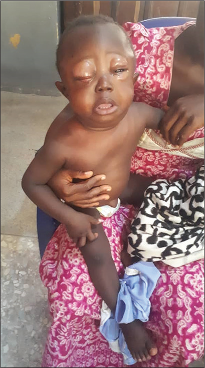
- Clinical manifestations at the time of admission.
An assessment of bronchopneumonia complicated by extensive subcutaneous emphysema was made. He was commenced on intravenous Gentamycin in addition to Amoxicillin-Clavulanate and was also administered an anxiolytic (chlorpheniramine).
Results of complete blood count and serum electrolytes were all within normal limits. His HIV screening was negative and blood culture yielded no growth. A babygram [Figure 2] and skull X-ray [Figure 3] of the child showed streaky areas of lucency under the skin of the scalp, neck, trunk, and upper limbs with right-sided pneumothorax.
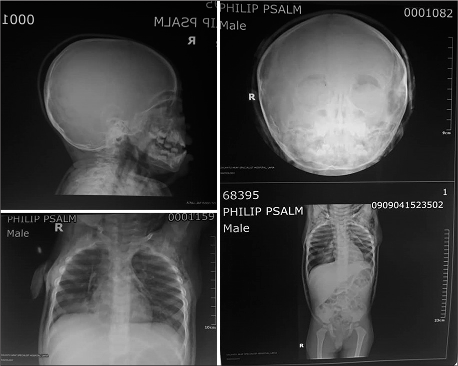
- Babygram showing air trapping under the skin of skull, neck and thorax.
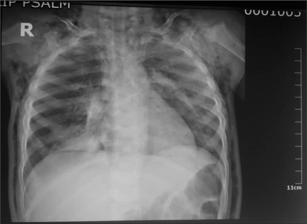
- CXR showing minimal pneumothorax.
Resolution of emphysema was noticeable on the 3rd day of admission and was completed by the 5th day. The baby’s vital signs remained stable, abnormal chest signs also resolved and the child was subsequently discharged after 7 days of hospitalization. The baby has been seen thrice on follow-up and has been healthy.
DISCUSSION
Subcutaneous emphysema results from processes allowing free air to enter into the subcutaneous tissue.[1] It is most commonly caused by pneumomediastinum or pneumothorax but rarely, air may be formed in the subcutaneous tissues by gas-forming bacteria.[1] The primary diagnosis of the index case is pneumonia, which was complicated by SSE and pneumothorax. This is consistent with the report by Okposio and Unior at a Central Hospital in Delta State.[11]
Therapeutic response to antibiotics would suggest a bacterial etiology of pneumonia in the highlighted case, although the full blood count was normal and blood culture was negative for any bacterial pathogen. This is consistent with earlier reports.[3,7] Okposio and Unior,[11] however, found elevated white cell count in their report. The index case had received 5 days of antibiotics before presentation with resolved pyrexia and this may have accounted for the absence of leukocytosis and negative blood culture.
The extensiveness of subcutaneous emphysema, in this case, is similar to the reports from a retrospective review by Abril and Ocete[9] in Granada, Spain, and this is due to the continuum of fascia plane across the body. The evolution of the emphysema could also have been aggravated by the high-flow nasal cannula (HFNC) oxygen therapy he had received from the referral health facility. Although HFNC is a safe method of effective oxygen delivery in children, it could be complicated by barotrauma such as pneumothorax and pneumomediastinum.[12]
Mild anxiolytic was administered to the patient to minimize the anxiety and restlessness (manifesting as excessive crying) as these could increase airway pressure and worsen emphysema.[5,9] The patient recovered fully on conservative care and was discharged on the 7th day on admission. This is similar to earlier reports by Siddki and Agrawad[13] in the United Kingdom, Akinyemi et al.[7] and Okposio and Unior[11] in Abeokuta and Delta in Nigeria, respectively. However, Aliyu[5] in Kano Nigeria discharged after 2 weeks on admission due to prolong oxygen therapy reflecting the severity of the underlying disease.
CONCLUSION
SSE associated with pneumothorax is a rare complication of childhood pneumonia but should be anticipated among subjects receiving high-flow oxygen therapy. Management is largely conservative and directed toward the cause.
Acknowledgment
We acknowledged the parent of the child for consenting and members of the health-care team for their efforts in the child’s care.
Declaration of patient consent
The authors certify that they have obtained all appropriate patient consent.
Financial support and sponsorship
Nil.
Conflicts of interest
There are no conflicts of interest.
References
- Emphysema and over-inflation In: Kliegman RM, Jenson HB, Behrman RE, Stanton BF, eds. Nelson Textbook of Paediatrics (18th ed). Philadelphia, PA: Saunders Elsevier; 2017. p. :1778-80.
- [Google Scholar]
- Massive spontaneous subcutaneous emphysema. Am J Med. 2016;129:e337-8.
- [CrossRef] [PubMed] [Google Scholar]
- Spontaneous subcutaneous emphysema and pneumomediastinum in a child with bronchial asthma. Ann Niger Med. 2015;9:30-2.
- [CrossRef] [Google Scholar]
- Tube thoracostomy: Complications and its management. Pulm Med. 2012;2012:256878.
- [CrossRef] [PubMed] [Google Scholar]
- Air leak syndrome complicating measles: Report of two cases. Menoufa Med J. 2016;29:174-6.
- [CrossRef] [Google Scholar]
- Subcutaneous emphysema of scalp following resuscitation in a neonate. J Trop Pediatr. 2017;63:499-501.
- [CrossRef] [PubMed] [Google Scholar]
- Pneumomediastinum and subcutaneous emphysema complicating acute exacerbation of bronchial asthma. Ann Ib Postgrad Med. 2007;5:78-9.
- [CrossRef] [PubMed] [Google Scholar]
- Subcutaneous emphysema and pneumomediastinum in child with asthma revealing occult foreign body aspiration: A case report. J Med. 2019;13:157-60.
- [CrossRef] [PubMed] [Google Scholar]
- Subcutaneous emphysema in critically Ill children. J Clin Med Imaging. 2018;4:1020-4.
- [Google Scholar]
- Pneumomediastinum and subcutaneous cervical emphysema: Unusual complications of childhood pneumonia. Niger J Paediatr. 2014;41:81-3.
- [CrossRef] [Google Scholar]
- High-flow nasal cannula oxygen therapy in children : A clinical review. Clin Exp Pediatr. 2020;63:3-7.
- [CrossRef] [PubMed] [Google Scholar]
- Case 4: Spontaneous emphysema in a toddler. Paediatr Child Health. 2015;21:139-40.
- [CrossRef] [PubMed] [Google Scholar]


