
Translate this page into:
“H” type tracheoesophageal fistula associated with cardiac and vertebral defects: management challenges at our institute – A case report with literature review

*Corresponding author: Manoj Kumar Mohanty, Department of Pediatric Surgery, All India Institute of Medical Sciences, Bhubaneswar, Odisha, India. pedsurg_manoj@aiimsbhubaneswar.edu.in
-
Received: ,
Accepted: ,
How to cite this article: Narahari J, Manekar AA, Tripathy BB, Mohanty MK. “H” type tracheoesophageal fistula associated with cardiac and vertebral defects: management challenges at our institute – A case report with literature review. J Pan Afr Thorac Soc. doi: 10.25259/JPATS_29_2024
Abstract
The “H” type tracheoesophageal fistula (H-TEF) is a rare, isolated congenital anomaly with an incidence of 4–5%, and it is rarely associated with other congenital conditions. Both diagnosis and surgical identification of this condition can be challenging. In most reported cases, the diagnosis was based on clinical suspicion. We present the case of a 1-year and 9-month-old male child who had regurgitation of feeds from the nose and mouth since birth, coughing after each feed, and failure to thrive. Additionally, the child had a small ventricular septal defect and gait abnormalities, including scoliosis due to butterfly vertebrae. The diagnosis was made through clinical presentation, a high index of suspicion, and fiberoptic bronchoscopy. Surgical management involved cannulating the fistula before ligation via a transcervical approach, though the initial attempt at cannulation was unsuccessful. A review of the literature is included, focusing on the clinical presentation of H-TEF, diagnostic methods, and treatment options. Early diagnosis and treatment of H-TEF can lead to symptom resolution, weight gain, and improved respiratory function.
Keywords
Butterfly vertebra
Flexible bronchoscopy
H-type tracheoesophageal fistula
Pediatric age
Recurrent respiratory tract infections

INTRODUCTION
H-type tracheoesophageal fistula (H-TEF) is a rare and life-threatening congenital anomaly characterized by an abnormal connection between the esophagus and trachea, accounting for 4–5% of all cases of esophageal atresia/tracheoesophageal fistula (EA/TEF).[1,2] It was first described by Lamb in 1873. H-TEF develops due to the failure of fusion of the tracheoesophageal ridges during the 3rd week of embryological development.[3] Common clinical manifestations include recurrent respiratory infections, aspiration during feeding with cyanosis, and abdominal distension. Diagnosing H-TEF can be challenging, often remaining undetected until later infancy or childhood. In our case, the diagnosis was made in infancy through high clinical suspicion. The first surgical repair of H-TEF was reported by Imperatori in 1939.[4] Here, we present an intriguing case of H-TEF diagnosed at the age of 1 year and 9 months.
CASE REPORT
A 1-year and 9-month-old male child presented with regurgitation of feeds through the nose and mouth after each feeding, along with coughing and failure to thrive since birth. He also exhibited an abnormal gait. Despite multiple hospital admissions, conservative management was applied each time. Chest X-ray lung field appeared normal, while a 2-D echocardiogram showed a small ventricular septal defect (VSD) measuring 7 mm. X-ray of the child revealed a butterfly vertebra at the T10 level [Figure 1]. A contrast-enhanced computed tomography scan of the thorax and neck showed air space consolidation in the right upper lobe and the posterior basal segments of both lower lobes, but no apparent TEF. A small soft tissue density (10 mm × 12 mm) with mild luminal narrowing was noted in the mid-esophagus. Given the recurrent upper respiratory tract infections, repeated hospital admissions, and failure to thrive, H-TEF was suspected, and the child was referred to our institute for further evaluation.
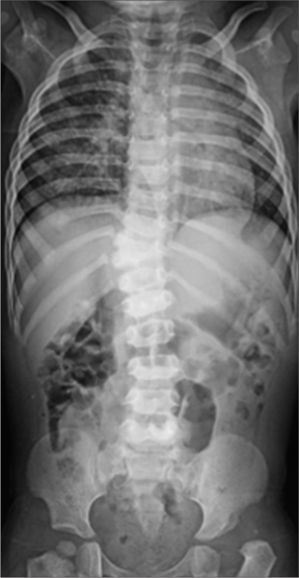
- Butterfly vertebra at T10 level.
Initially, the child was managed with nasogastric (NG) feeds, nebulization with normal saline, and oral antibiotics via the NG tube. Surgery was planned, and preoperative fiberoptic bronchoscopy revealed a fistulous opening at the 6 o’clock position in the trachea, 2.5 cm above the carina, which appeared large. Attempts to cannulate the fistula using a 4 French (FR) Fogarty catheter were unsuccessful after 2–3 attempts, causing laryngeal edema, and the procedure was abandoned. The child was then managed in the intensive care unit with steroids and intravenous (IV) antibiotics.
Ten days later, the child was re-operated, and the fistula was successfully cannulated using the 4 FR Fogarty catheter. The right lower horizontal cervical incision was given. Identification of the trachea and esophagus was done followed by identification of fistula located at the level of T1 vertebra [Figure 2]. Ligation and repair of the fistula with meticulous closure at the tracheal and esophageal end was done. The neck wound was closed keeping a suction drain. The procedure was uneventful. Postoperatively, the child was kept nil per oral for 5 days, received IV fluids and antibiotics, and a contrast study on the 5th postoperative day (POD) showed no leakage. The NG tube was removed on the 7th POD, and oral feeds were reintroduced. There was no regurgitation or coughing after feeds. The child was discharged on the 10th POD in stable condition.
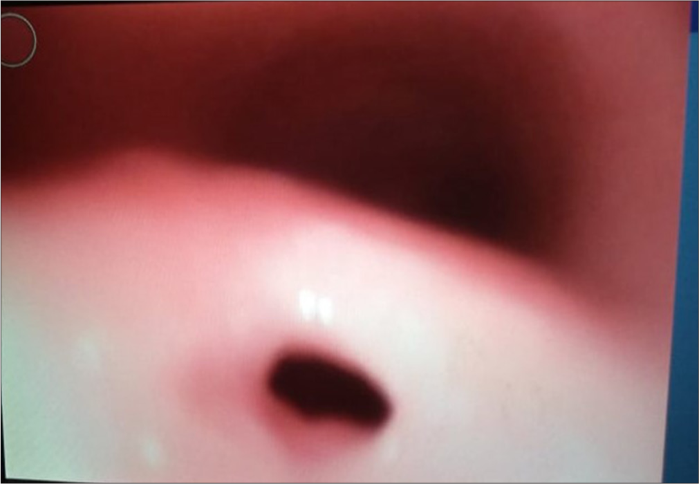
- Flexible bronchoscopy depicting H-type tracheoesophageal fistula just above the carina.
DISCUSSION
H-TEF accounts for 4–5% of all congenital tracheoesophageal malformations, with an incidence ranging from 1.8% to 4.2%.[5] The classical symptoms of H-TEF include recurrent chest infections, choking during feeding, and failure to thrive. In our case, these symptoms were present, along with an abnormal gait. The most prominent symptom is regurgitation and choking during feeding, typically starting from birth, although severity and frequency may vary.
H-TEF is the least associated with other congenital anomalies, occurring in about 30% of cases. In contrast, other variants of esophageal atresia with TEF are more commonly associated with multiple anomalies. Commonly associated abnormalities include VACTERL/VATER association, VSD, vascular ring, Fallot’s tetralogy, chromosomal anomalies, duodenal atresia, renal anomalies, vertebral anomalies, CHARGE association, Goldenhar’s syndrome, syndactyly, esophageal stenosis, laryngeal cleft, malrotation, imperforate anus, and Trisomy 21.[6] Our case involved a vertebral anomaly (butterfly vertebra) and a congenital cardiac anomaly (7 mm VSD). Early recognition of symptom complexes and a high clinical index of suspicion are essential for early diagnosis. Killen and Greenlee reported that 43% of diagnoses are made within the 1st month of life, and 83% within the 1st year. Delayed diagnosis, due to failure to identify the fistula or improper radiological techniques, may lead to fatal consequences.
The diagnosis of H-TEF requires confirmation of the presence of a fistula and its precise location. Chest radiography, while not diagnostic, can be helpful, often showing aspiration pneumonitis and gastric distension, which raise suspicion for H-TEF, especially in infants with suggestive clinical signs.
Rigid bronchoscopy is considered the most reliable diagnostic method due to superior optics and the ability to gently probe the posterior tracheal wall, which may reveal the fistula opening. Cannulating the fistula helps minimize gastric distension and its detrimental effects on ventilation, facilitating quick identification and surgical preparation.[7] Many surgeons now recommend the use of preoperative or intraoperative bronchoscopic guidewire transfistula placement.[8] This technique offers several advantages. First, the wire can be passed through a flexible bronchoscope, and its position can be confirmed on a chest X-ray, ensuring the fistula’s exact location is identified. Second, the wire can be easily palpated during surgery, aiding in the precise identification of the fistula and helping preserve surrounding structures. In place of a vascular guidewire, Fogarty or ureteral catheters can also be used as alternatives. In our case, the fistula was initially unsuccessful in cannulating with a Fogarty catheter, but it was successfully cannulated after 10 days.
Bronchoscopy serves several purposes: Identifying the fistula’s level, detecting unusual variants (such as double or trifurcation fistulas), assessing tracheobronchitis severity, determining the aortic arch position, and confirming the placement of a Fogarty catheter. Bronchoscopy appears safe and beneficial for all infants with esophageal atresia and TEF.[9]
The primary goal in treating H-TEF is the closure of the fistula, which is most effectively achieved through surgical division. Surgical approaches vary, but cervicotomy is typically preferred for proximally located fistulas, while thoracotomy is used for more distally located fistulas.[4] Cervicotomy is associated with lower postoperative morbidity compared to thoracotomy. Brookes et al. reported that fistulas located between C5 and T3 were successfully divided through a right cervicotomy.[10] Sundar et al. achieved similar results with a left cervical approach. In our case, the fistula was accessed via a right cervicotomy, with the fistula located at the T1 vertebra.[11] Despite cannulating the fistula, we encountered difficulty ligating it due to its low position.
Endoscopic methods for fistula closure, such as tissue adhesives, electrocautery, sclerosants, and lasers, have been reported. Bhatnagar et al. found endoscopic treatment of recurrent and congenital H-TEF easier than open surgery, offering a safer alternative.[12] Neodymium-doped yttrium aluminum garnet lasers were noted to be more effective than electrocautery for obliterating the fistula. Other techniques, including electrocautery and histoacryl glue, have been successful in managing H-type and recurrent TEFs. However, if a fistula does not close after two endoscopic attempts, it is recommended to proceed with an external surgical approach.[13]
CONCLUSION
H-TEF is a rare condition with a more favorable prognosis when diagnosed early. A high clinical index of suspicion and recognition of classic symptoms is essential for accurate diagnosis. H-TEF is associated with the least number of congenital anomalies, occurring in approximately 30% of cases. However, in our case, the child presented with vertebral defects (butterfly vertebra) and cardiac anomalies (VSD). Cannulating the fistula before ligation can be challenging, and multiple attempts should be avoided to prevent edema and minimize the risk of injury to the esophagus.
Ethical approval
Institutional Review Board approval is not required.
Declaration of patient consent
The authors certify that they have obtained all appropriate patient consent.
Conflicts of interest
There are no conflicts of interest.
Use of artificial intelligence (AI)-assisted technology for manuscript preparation
The authors confirm that there was no use of artificial intelligence (AI)-assisted technology for assisting in the writing or editing of the manuscript and no images were manipulated using AI.
Financial support and sponsorship: Nil.
References
- Etiology of esophageal atresia and tracheoesophageal fistula: “Mind the gap”. Curr Gastroenterol Rep. 2010;12:215-22.
- [CrossRef] [PubMed] [Google Scholar]
- The epidemiology of tracheooesophageal fistula and oesophageal atresia in Europe. EUROCAT Working Group. Arch Dis Child. 1993;68:743-8.
- [CrossRef] [PubMed] [Google Scholar]
- Esophageal atresia and tracheoesophageal fistula. Am Fam Physician. 1999;59:919-20.
- [Google Scholar]
- 'H' type tracheooesophageal fistula-case reports with review of the literature. Egypt J Ear Nose Throat Allied Sci. 2014;15:143-8.
- [CrossRef] [Google Scholar]
- The diagnosis of congenital tracheooesophageal fistula. J Pediatr Surg. 1988;23:415-7.
- [CrossRef] [PubMed] [Google Scholar]
- Bronchoscopy in newborns with esophageal atresia. Pediatr Med Chir. 2002;24:297-301.
- [Google Scholar]
- Flexible bronchoscopic cannulation of an isolated H-type tracheoesophageal fistula in a newborn. J Pediatr Surg. 2012;47:e9-10.
- [CrossRef] [PubMed] [Google Scholar]
- Crucial bronchoscopic findings in esophageal atresia and tracheoesophageal fistula. J Pediatr Surg. 1988;23:466-70.
- [CrossRef] [PubMed] [Google Scholar]
- H-type congenital tracheoesophageal fistula: University of Iowa experience 1985 to 2005. Ann Otol Rhinol Laryngol. 2007;116:363-8.
- [CrossRef] [PubMed] [Google Scholar]
- Congential H-type tracheooesophageal fistula. Arch Dis Childhood. 1975;50:862-3.
- [CrossRef] [PubMed] [Google Scholar]
- Endoscopic treatment of tracheoesophageal fistula using electrocautery and the Nd: YAG laser. J Pediatr Surg. 1999;34:464-67.
- [CrossRef] [PubMed] [Google Scholar]
- Endoscopic treatment of congenital H-Type and recurrent tracheoesophageal fistula with electrocautery and histoacryl glue. Int J Pediatr Otorhinolaryngol. 2006;70:925-30.
- [CrossRef] [PubMed] [Google Scholar]


